
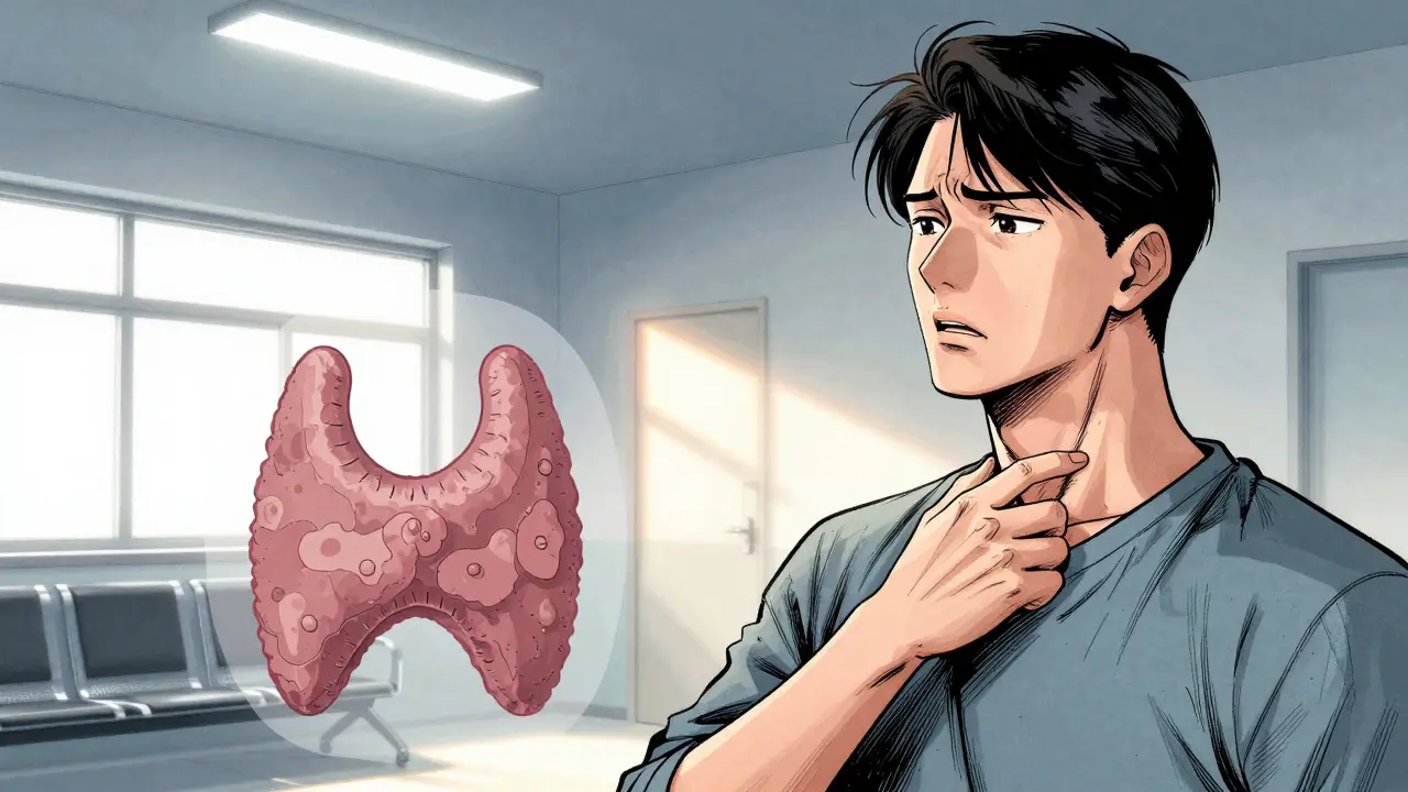
Imagine finding a lump in your neck. Your heart races. The internet tells you it’s cancer. But here is the truth that might calm your nerves: most lumps on the thyroid gland are not dangerous. Thyroid nodules are incredibly common. In fact, if you get an ultrasound after age 60, there is a two-in-three chance one will show up. Yet, only about 5% to 10% of these nodules turn out to be malignant. The real challenge isn't just finding them-it's knowing which ones need action and which ones can safely be ignored.
Knowing when to worry and when to wait saves you from unnecessary surgeries and anxiety. This guide breaks down how doctors tell the difference between harmless growths and those that require a thyroid biopsy, using the latest medical standards so you can understand your own diagnosis with confidence.
What exactly is a thyroid nodule?
A thyroid nodule is a discrete lump or lesion within the thyroid gland that feels different from the surrounding tissue. Most are fluid-filled cysts or solid collections of thyroid cells. They are usually detected via ultrasound or physical exam and are often asymptomatic.
The Silent Growth: Why Size and Speed Matter
You cannot judge a nodule by its size alone. A small nodule can be aggressive, while a large one might be perfectly benign. However, size does set the threshold for investigation. According to guidelines from the American Thyroid Association, any nodule larger than 1 centimeter (about the size of a pea) warrants closer look. But the real red flag isn't just how big it is-it's how fast it grows.
Research published in the Journal of Clinical Endocrinology & Metabolism highlights a critical metric: growth rate. If a nodule grows more than 2 millimeters per year, the risk of malignancy jumps significantly. For every additional 2 mm/year above that threshold, the danger increases further. In contrast, benign nodules typically grow at a sluggish pace of about 1 mm per year. This distinction is why doctors insist on serial ultrasounds. They aren't just measuring; they are timing.
- Benign Growth: Slow, steady increase (~1 mm/year). Often stable over years.
- Suspicious Growth: Rapid expansion (>2 mm/year). Requires immediate re-evaluation.
- Stable Nodules: No change in size over 24 months. Usually safe for continued monitoring.
If your doctor measures your nodule today and again in six months, they are looking for this velocity. A sudden jump in size triggers the next step: determining what the nodule looks like under the hood.
Ultrasound Clues: What Doctors Look For
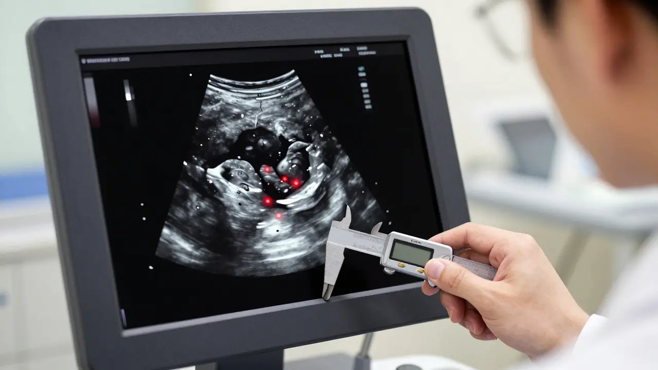
An ultrasound is the detective’s magnifying glass. It doesn't just show size; it reveals texture and structure. Radiologists use specific features to assign a risk score, often using systems like TI-RADS (Thyroid Imaging Reporting and Data System). Not all nodules look the same, and their appearance tells a story.
Malignant nodules tend to have "ugly" features. They are often hypoechoic, meaning they appear darker than the surrounding thyroid tissue because they are denser. They may have irregular margins-jagged edges that suggest the nodule is invading nearby tissue rather than staying contained. Perhaps the most concerning sign is microcalcifications. These tiny bright spots inside the nodule are present in over half of papillary carcinomas, the most common type of thyroid cancer.
On the flip side, benign nodules often look "pretty." They might be spongiform, resembling a sponge with many small cystic spaces. Pure cysts, which are entirely fluid-filled, are almost always benign. If your ultrasound report mentions "spongiform" or "cystic," take a deep breath. Those are good words.
| Feature | Benind Likelihood | Suspicious/Malignant Likelihood |
|---|---|---|
| Echogenicity | Isoechoic or Hyperechoic (bright) | Hypoechoic (dark) |
| Margins | Smooth, well-defined | Irregular, infiltrative |
| Composition | Cystic or Spongiform | Solid |
| Calcifications | None or coarse macrocalcifications | Microcalcifications (tiny bright spots) |
| Shape | Taller-than-wide (less common) | Taller-than-wide (vertical orientation) |
The Bethesda System: Decoding Your Biopsy Results
If your nodule has suspicious features and is large enough, your doctor will recommend a fine-needle aspiration (FNA) biopsy. This procedure involves inserting a thin needle into the nodule to extract cells. The results don't come back as simple "yes" or "no." Instead, they use the Bethesda System for Reporting Thyroid Cytopathology, a standardized classification system created to reduce confusion and guide treatment.
Understanding your Bethesda category is crucial because it dictates your next move. Here is what each category means for you:
- Category I (Nondiagnostic): Not enough cells were collected. Risk of cancer is low (1-4%), but you’ll likely need a repeat biopsy.
- Category II (Benign): The cells look normal. Risk is near zero (0-3%). You can usually stop worrying and just monitor with ultrasound.
- Category III (Atypia of Undetermined Significance): The cells look slightly weird but not clearly cancerous. Risk is 5-15%. This is where molecular testing helps decide if surgery is needed.
- Category IV (Follicular Neoplasm): Cells suggest a tumor, but we can't tell if it's invasive without seeing the whole capsule. Risk is 15-30%. Often requires diagnostic surgery.
- Category V (Suspicious for Malignancy): High suspicion. Risk is 60-75%. Surgery is typically recommended.
- Category VI (Malignant): Cancer is confirmed. Risk is 97-99%. Surgical removal is the standard next step.
Categories III and IV are the "gray zones." Historically, patients in these groups faced unnecessary surgeries because doctors couldn't be sure. Today, molecular testing changes the game.
Molecular Testing: Cutting Through the Gray Zone
When your biopsy result is indeterminate (Bethesda III or IV), you face a dilemma: operate and risk removing a healthy gland, or wait and risk missing a cancer? Enter molecular testing. Tests like Afirma GSC or ThyroSeq v3 analyze the genetic material of the nodule cells.
Think of it as checking the engine code of a car. Even if the car looks fine on the outside (benign cytology), the computer might reveal a faulty sensor (genetic mutation). Afirma has a negative predictive value of 97%, meaning if it says "benign," you can trust it. ThyroSeq v3 analyzes 112 genes with high sensitivity. These tests have reduced unnecessary surgeries by up to 35% in recent studies. If your doctor suggests molecular testing after an indeterminate biopsy, it’s a sign they want to avoid cutting unless absolutely necessary.

Who Needs a Biopsy? The Decision Rules
Not every nodule gets biopsied. Over-biopsying leads to false positives and unnecessary stress. The American Thyroid Association provides clear rules based on size and ultrasound features. Here is a simplified breakdown of when a biopsy is typically needed:
- Nodules ≥ 1 cm with suspicious features: Biopsy is recommended. Suspicious features include hypoechogenicity, microcalcifications, or irregular borders.
- Nodules ≥ 1.5 cm without suspicious features: Biopsy may be considered, especially if there is a family history of thyroid cancer or prior radiation exposure.
- Nodules ≥ 2 cm regardless of features: Biopsy is generally recommended due to the sheer size, even if it looks benign, to rule out rare aggressive types.
- Rapid Growth: Any nodule growing >2 mm/year should be biopsied, regardless of initial appearance.
If you have a history of head and neck radiation or a family history of medullary thyroid cancer or multiple endocrine neoplasia (MEN), the thresholds drop lower. In these cases, even smaller nodules warrant attention.
Living With a Nodule: Monitoring and Management
For the vast majority of people, a thyroid nodule is a spectator sport. You watch it, but it doesn't play a role in your daily life. Active surveillance is the standard for benign nodules. This means getting an ultrasound every 6 to 12 months initially, then annually if stable. If the nodule stays the same size for two years, the interval can often be extended.
Surgery is reserved for nodules that are cancerous, highly suspicious, or causing physical problems. Large benign nodules can sometimes press on the windpipe or esophagus, causing trouble breathing or swallowing. In these cases, procedures like radiofrequency ablation (RFA) are emerging as alternatives to surgery. RFA shrinks the nodule by heating it, offering volume reduction with fewer complications than traditional thyroidectomy. Recent trials show RFA can reduce nodule volume by 78% at 12 months with a much lower complication rate than surgery.
Remember, thyroid cancer, particularly papillary carcinoma, is often slow-growing. Even if diagnosed, the prognosis is excellent. The goal of modern medicine is not just to find cancer, but to find the right cancer to treat, sparing you from overtreatment. Trust the data, follow the guidelines, and partner with your endocrinologist to make decisions that fit your specific health profile.
Is a thyroid biopsy painful?
Most patients describe the discomfort as mild. The area is numbed with local anesthesia before the needle is inserted. You may feel pressure or a slight pinch, but severe pain is uncommon. The procedure typically takes less than 15 minutes.
Can thyroid nodules go away on their own?
Solid nodules rarely disappear completely. However, cystic nodules (fluid-filled) can shrink or resolve spontaneously. Some nodules also stabilize and stop growing, effectively becoming inactive. Regular monitoring ensures that any changes are caught early.
Do I need to change my diet if I have a thyroid nodule?
There is no specific "nodule diet." However, maintaining adequate iodine intake is important for overall thyroid health. Avoid excessive consumption of goitrogens (like raw cruciferous vegetables) only if you have an iodine deficiency. For most people, a balanced diet is sufficient. Always consult your doctor before taking supplements.
What causes thyroid nodules?
The exact cause is often unknown. Factors include iodine deficiency, genetic mutations, inflammation (thyroiditis), and aging. Women are three times more likely to develop nodules than men. Radiation exposure to the head and neck area is a known risk factor for both benign and malignant nodules.
How accurate is an ultrasound in detecting thyroid cancer?
Ultrasound is highly sensitive, with detection rates around 87-94% when combined with size criteria. However, it cannot definitively diagnose cancer; it only identifies suspicious features. A biopsy is required for a definitive diagnosis. Ultrasound remains the gold standard for initial evaluation and monitoring.