

Imagine finding out you have cancer. The immediate question isn't just "what kind?" but "where is it exactly, and how far has it spread?" This is where oncologic imaging steps in. It’s not just about taking pictures; it’s about mapping the battlefield so doctors can fight effectively. For decades, we relied on basic X-rays or ultrasounds, but today, the gold standards are PET-CT, a hybrid imaging technique combining Positron Emission Tomography and Computed Tomography to show metabolic activity and anatomical structure simultaneously, MRI, Magnetic Resonance Imaging, which uses strong magnetic fields and radio waves to create detailed images of organs and tissues without ionizing radiation, and the newer PET-MRI, an advanced hybrid system that combines the functional data of PET with the superior soft-tissue contrast of MRI in a single scan.
Choosing the right tool matters. A wrong stage means a wrong treatment plan. In this guide, we break down how these technologies work, their specific strengths in staging accuracy, and when one beats the other. We’ll look at real-world performance in cancers like breast, prostate, and lung, helping you understand what your medical team is considering.
How These Imaging Tools Actually Work
To understand why one might be better than another, you need to know what they’re actually measuring. They don’t just see shape; they see function and biology.
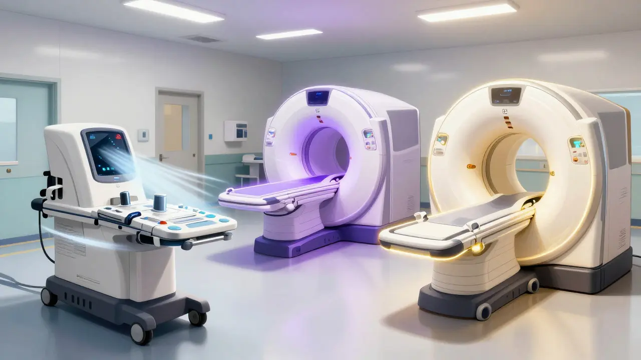
PET-CT was introduced clinically around 2001. It works by injecting a radioactive tracer, usually 18F-FDG, Fluorodeoxyglucose, a radioactive sugar analog used in PET scans to highlight areas of high metabolic activity like tumors. Cancer cells eat sugar faster than normal cells, so they light up on the PET scan. The CT part provides the map-the anatomy-so doctors know exactly where those bright spots are. It’s fast, taking about 15-20 minutes per patient, but it exposes you to radiation (10-25 mSv).
MRI, developed independently by Nobel laureates Paul Lauterbur and Peter Mansfield, uses magnets, not radiation. It’s incredible for seeing soft tissue details. If you’re looking at the brain, spine, or pelvic organs, MRI gives you crystal-clear resolution (down to 0.5-1.0 mm). However, it takes longer-30 to 60 minutes-and if you have metal implants, you might not even be able to use it.
PET-MRI is the new kid on the block, commercially available since 2010 via systems like the Siemens Biograph mMR. It tries to give you the best of both worlds: the metabolic heat map from PET and the high-definition soft tissue detail from MRI, all in one session. It cuts radiation exposure by about 50% compared to PET-CT because it skips the CT component. But it’s slower, more expensive, and technically trickier to run.
Staging Accuracy: Who Wins?
"Staging" determines the size of the tumor and whether it has spread to lymph nodes or other organs. Accuracy here is life-or-death. Let’s look at how these modalities stack up in specific scenarios.
| Cancer Type / Scenario | Best Modality | Key Metric / Finding | Why It Wins |
|---|---|---|---|
| Breast Cancer (Response to Chemo) | PET-CT | Specificity: 0.94 vs 0.83 for MRI | Better at distinguishing active tumor from inflammation early in treatment. |
| Pancreatic Cancer | PET-MRI | Changed management in 49% of cases | Sensitive to small lesions in complex abdominal anatomy. |
| Prostate Cancer | MRI (Conventional) | Accuracy: 75% vs 62% for PSMA PET/CT | Superior local tissue definition for primary tumor assessment. |
| Lung Cancer (Lymph Nodes) | PET-CT & PET-MRI (Tie) | Diagnostic Accuracy: 0.84 for both | Both effectively detect metabolic activity in nodes. |
| Brain Tumors (Recurrence vs. Necrosis) | PET-MRI | Accuracy: 85-90% vs 70-80% for MRI alone | Critical distinction between live tumor and radiation damage. |
In breast cancer, for example, a 2017 study in *Nature Scientific Reports* showed that PET-CT had higher specificity (0.94) than conventional contrast-enhanced MRI (0.83) when evaluating response to neoadjuvant chemotherapy before three cycles were complete. Why? Because MRI can sometimes mistake inflammation for tumor, while PET looks at actual cellular energy use.
However, for pancreatic cancer, the story flips. A 2023 study in *RadioGraphics* found that FDG PET/MRI changed management decisions for nearly half (49%) of patients compared to standard protocols. The pancreas is tucked away behind other organs, making it hard to see. The combined sensitivity of PET-MRI helps spot tiny metastases that others miss.
For prostate cancer, it’s complicated. While PSMA PET/CT is gaining popularity, a 2022 publication in *Cancer* journal noted that conventional MRI still detects prostate cancer with 75% accuracy, outperforming PSMA PET/CT’s 62% in certain contexts. This highlights that no single test is perfect for every job.
The Practical Reality: Cost, Time, and Access
If PET-MRI is so good, why isn’t everyone using it? The answer lies in logistics and economics.
Cost: According to 2023 US pricing, a PET-MRI exam costs approximately $2,500-$3,500, whereas a PET-CT runs $1,600-$2,300. That’s a 50% premium. Dr. Barry Siegel from Washington University School of Medicine noted in a 2020 *JAMA Oncology* commentary that this higher cost requires careful patient selection. Hospitals aren’t going to order the most expensive test unless there’s a clear diagnostic advantage.
Time: PET-CT is fast. You’re in and out in under 20 minutes of scanning time. MRI takes 30-60 minutes. PET-MRI? Also 45-60 minutes. For anxious patients, or those who are frail, lying still in a narrow tube for an hour is tough. Motion artifacts can ruin the image, as Dr. Sarah Chen from Massachusetts General Hospital pointed out in a 2023 ACR forum discussion.
Availability: PET-CT is the "workhorse." It’s widely available in community hospitals. PET-MRI is mostly found in academic medical centers. A 2022 HIMSS Analytics report stated that 78% of PET-MRI installations are in institutions with residency training programs. If you’re in a rural area, you likely won’t have access to PET-MRI.
Expert Consensus: When to Use What
Radiologists don’t pick tests based on hype; they pick them based on evidence. Here’s what the experts say:
- Dr. Richard L. Wahl (Johns Hopkins): He calls PET-MRI "particularly valuable for pelvic malignancies and CNS tumors" due to its soft-tissue contrast. But he admits PET-CT remains the standard for most indications because it’s faster and more available.
- EANM/ESMRMB Consensus (2019): This major European guideline concluded that PET-MRI offers "moderate advantages" for liver metastases, pelvic cancers, and pediatric cases. Crucially, they said it does not represent a universal replacement for PET-CT.
- Dr. Hedvig Hricak (Memorial Sloan Kettering): She emphasizes personalization. "No single imaging method is optimal for all cancer scenarios," she stated in 2021. The choice depends on tumor biology, the specific clinical question, and available resources.
The trend is moving toward precision. The 2024 ASCO guidelines now recommend selecting imaging based on tumor molecular subtypes. For instance, in brain tumors, differentiating between tumor recurrence and radiation necrosis is critical. MRI alone gets it right 70-80% of the time. PET-MRI boosts that to 85-90%, potentially sparing patients from unnecessary biopsies or surgeries.
Future Trends: AI and New Tracers
We’re entering an era of convergence. At the 2023 RSNA annual meeting, there were 27 presentations on AI-enhanced quantitative imaging biomarkers. Artificial intelligence is starting to read these scans, predicting treatment responses before they’re visible to the human eye.
New tracers are also changing the game. 68Ga-PSMA-11, A radiotracer targeting Prostate-Specific Membrane Antigen, FDA-approved in 2020 for detecting prostate cancer recurrence is revolutionizing prostate cancer care. Combining this with multiparametric MRI could offer unprecedented clarity.
Hardware is improving too. Siemens Healthineers announced the BioMatrix 600 PET/MRI in January 2024, featuring a wider bore and a 6-minute whole-body acquisition time. This addresses the biggest complaint about PET-MRI: speed. By 2035, analysts predict PET-MRI will capture 25-30% of the market in academic centers, driven by the push to reduce radiation doses, especially in younger patients.
Conclusion: There Is No One-Size-Fits-All
Oncologic imaging is not a competition with a single winner. It’s a toolbox. PET-CT is reliable, fast, and widely available. MRI offers stunning detail without radiation. PET-MRI is the high-precision instrument for complex cases where every millimeter counts.
As a patient, ask your doctor: "What specific question are we trying to answer with this scan?" If it’s initial staging of lung cancer, PET-CT is likely sufficient. If it’s checking for recurrence in the brain after radiation, PET-MRI might be worth the extra cost and time. Understanding these tools empowers you to participate in your care decisions with confidence.
Is PET-MRI better than PET-CT for all types of cancer?
No. While PET-MRI offers superior soft-tissue contrast and lower radiation, PET-CT remains the standard for many cancers due to its speed, availability, and proven efficacy in lung and lymph node staging. PET-MRI is particularly advantageous for brain, pelvic, and pediatric cancers, but it is not a universal replacement.
Which imaging modality has the least radiation exposure?
MRI has zero ionizing radiation. PET-MRI reduces radiation exposure by approximately 50% compared to PET-CT because it eliminates the CT component's radiation dose. Standard PET-CT exposes patients to 10-25 mSv of radiation, depending on the protocol.
Why is PET-MRI more expensive than PET-CT?
PET-MRI systems cost significantly more to purchase ($3.0-$4.2 million vs. $1.8-$2.5 million for PET-CT) and require specialized infrastructure like RF-shielded rooms. Additionally, the longer scan times (45-60 minutes) reduce patient throughput, increasing operational costs per examination.
Can MRI detect cancer as well as PET-CT?
It depends on the cancer type. MRI excels at visualizing soft tissue structures in the brain, pelvis, and breasts. However, PET-CT is often better at detecting distant metastases and assessing overall metabolic activity throughout the body. For some cancers, like prostate, MRI may have higher local accuracy, while PET-CT is better for systemic staging.
How long does a PET-MRI scan take?
A PET-MRI scan typically takes 45 to 60 minutes. This is longer than a PET-CT scan (15-20 minutes) but similar to a comprehensive MRI exam. The longer duration can be challenging for patients who have difficulty remaining still, potentially leading to motion artifacts.